|
 |
Page d'accueil Actions engagees Actions engagees Axe diagnostique Axe diagnostique Neurobiologie Neurobiologie
|
 |
 |
Sonde TEP |
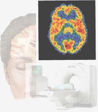
Une sonde biologique est en cours de conception pour l’exploration par tomographie cérébrale du système glutaminergique. La validation future de cette sonde sera un tournant décisif dans la prise en charge diagnostique et thérapeutique en neuropsychiatrie (notamment, concernant la schizophrénie et la maladie d’Alzheimer). |
 |
|
Communication |
23 janvier 2010
Le Dr Belhadj-Tahar a été invité lors du 37eme séminaire du Club Français des Radiopharmaceutiques qui s'est tenu le 22/01/2010 à la Faculté de Médecine Paul Sabatier de Toulouse, pour présenter les résultats des travaux de recherche concernant cette sonde TEP pour l'exploration cérébrale en vue de détection du stress neurotoxique. A cette occasion, le Dr Belhadj-Tahar a tenu à rappeler qu'actuellement il n'existe aucun traceur ou agent diagnostique dédié au dépistage précoce de la schizophrénie en particulier dont le pronostic dépend du délai de la mise en route du traitement adéquat dès la phase prodromique ( et qui peut durer de quelques mois à quelques années). De même il a conclu que la concrétisation de tel projet de NeuroPsychoPharmacologie nécessite une approche pluridisciplinaire intégrative en Chimie, Pharmacologie et imagerie. A ce titre l'AFPreMed a expérimenté avec succès ce concept d’interface intégrative. [Programme du 37eme séminaire du Club Français des Radiopharmaceutiques, Toulouse:22/01/2010 [249 KB]
] |
 |
|
Publication Scientifique |
07 avril 2015
Radiolabeling of [18F]-fluoroethylnormemantine and initial in vivo evaluation of this innovative PET tracer for imaging the PCP sites of NMDA receptors
Anne-Sophie Salabert [a, b, c], Caroline Fonta [c], Charlotte Fontan [a, b], Adel Djilali [a], Mathieu Tafani [a, b], Carine Pestourie [d], Hafid Belhadj-Tahar[e], Mathieu Alonso[b], Pierre Payoux[a, f]
a Brain Imaging and Neurological Disability UMR 825, INSERM, University of Toulouse UPS, Toulouse, France
b Radiopharmacy Department, University Hospital, Toulouse, France
c Brain and Cognition Research Center (CERCO), University of Toulouse UPS, CNRS-UMR 5549, Toulouse, France
d Animal experimentation platform, UMS 006, Toulouse, France
e Research and Expertise Group, French Association for the Promotion of Medical Research (AFPREMED), Toulouse, France
f Nuclear Medicine Department, University Hospital, Toulouse, France
Introduction
The N-methyl-D-aspartate receptor (NMDAr) is an ionotropic receptor that mediates excitatory transmission. NMDAr overexcitation is thought to be involved in neurological and neuropsychiatric disorders such as Alzheimer disease and schizophrenia. We synthesized [18 F]-fluoroethylnormemantine ([18 F]-FNM), a memantine derivative that binds to phencyclidine (PCP) sites within the NMDA channel pore. These sites are primarily accessible when the channel is in the active and open state.
Methods
Radiosynthesis was carried out using the Raytest® SynChrom R&D fluorination module. Affinity of this new compound was determined by competition assay. We ran a kinetic study in rats and computed a time-activity curve based on a volume-of-interest analysis, using CARIMAS® software. We performed an ex vivo autoradiography, exposing frozen rat brain sections to a phosphorscreen. Adjacent sections were used to detect NMDAr by immunohistochemistry with an anti-NR1 antibody. As a control of the specificity of our compound for NMDAr, we used a rat anesthetized with ketamine. Correlation analysis was performed with ImageJ software between signal of autoradiography and immunostaining.
Results
Fluorination yield was 10.5% (end of synthesis), with a mean activity of 3145 MBq and a specific activity above 355 GBq/μmol. Affinity assessment allowed us to determine [19F]-FNM IC50 at 6.1 10- 6 M . [18 F]-FMN concentration gradually increased in the brain, stabilizing at 40 minutes post injection. The brain-to-blood ratio was 6, and 0.4% of the injected dose was found in the brain. Combined ex vivo autoradiography and immunohistochemical staining demonstrated colocalization of NMDAr and [18 F]-FNM (r = 0.622, p < 0.0001). The highest intensity was found in the cortex and cerebellum, and the lowest in white matter. A low and homogeneous signal corresponding to unspecific binding was observed when PCP sites were blocked with ketamine.
Conclusions
[18F]-FNM appears to be a promising tracer for imaging NMDAr activity for undertaking preclinical studies in perspective of clinical detection of neurological or neuropsychological disorders
Keywords
activated NMDA receptor; innovative PET probe; preclinical; memantine; neuropathology; excitatory neurotransmission
[lire l'article] |
|
14/10/2014
Conception d'un nouveau traceur du système NMDA pour l‘exploration tomographique du stress neurotoxique.
Anne-Sophie BRUN-SALABERT
UMR 825 Inserm : imagerie cérébrale et handicap neurologique - Toulouse
Abstract :
Le récepteur N-méthyl-D-aspartate (NMDA) est un récepteur ionotropique qui assure la médiation de la transmission excitatrice. Sa suractivation entraîne une surexcitation neuronale et une excitotoxicité par un afflux prolongé de calcium. Ce processus est impliqué dans des troubles neurologiques (maladie d'Alzheimer) et neuropsychiatriques (état de stress post-traumatique, schizophrénie). A ce titre, le ligand fluoré (19F froid à l'échelle pondérale) antagoniste du récepteur NMDAr permet de modulation basse du système glutaminergique induisant un effet protecteur contre le stress neurotoxique et une action anxiolytique. Nous présentons les résultats de la radiosynthèse et de l'évaluation pharmacologique sur des rats d'une nouvelle sonde TEP au fluor (18FENM radioactif) dérivée de la mémantine pour I ‘exploration in vivo du NMDA. Cette étape de binding et localisation In vivo par tomographie TEP est indispensable pour déterminer l'utilisation de l'homologue fluoré froid (19FENM) de cette molécule pour notamment la neuroprotection et le stress post traumatique.
- CONGRES SCIENTIFIQUE “LA RECHERCHE EN PSYCHIATRIE: LES ENJEUX AU PRÉSENT ET À L'AVENIR” CONSACRE A : “ETAT DE STRESS POST TRAUMATIQUE”, Albi, le 14 Octobre 2014. |
|
 |
 |
-
|
|
Outil neurobiologique |
28/05/2009
L'équipe de neurobiologistes appartenant à l'AFPReMed travaille actuellement à la mise au point d'un outil pharmacologique permettant le diagnostic et le suivi des troubles neuropsychiatriques survenant notamment lors du syndrome de stress post-traumatique. L'aboutissement prochain de ce projet dépendra en partie de l'aide financière sollicitée auprès de généreux donateurs.
* Le démarrage de ce projet (avec le soutien de l’équipe EA3033) est prévu pour la fin du mois d’avril 2010.
03/05/2010
Les travaux de mise au point des analyses du BDNF (Brain-derived neurotrophic factor) dans le plasma et le liquide céphalo-rachidien ont commencé le 30/04/2010.
22/10/2014
Communication au 27eme congrès de ECNP le 18-21 octobre 2014 à Berlin:
"Brain-derived neurotrophic factor (BDNF) as a predictive factor for post-traumatic stress disorder (PTSD)"
[Lire l'abstract ]
|
 |
|
|
08/04/215
Publication des travaux concernant l'application du dosage de BDNF dans la detection précoces de l'état de stress Post traumatique:
Journal of US-China Medical Science 12 (2015) 12-19
doi: 10.17265/1548-6648/2015.01.002
Pilot Prospective Study on BDNF (Brain Derived Neurotrophic Factor) as a Predictive Biomarker of the Occurrence of PTSD (Post Traumatic Stress Disorder)
[Etude pilote prospective: la neurotrophine (BDNF) comme marqueur predictif de la survenue de l'état de stress post traumatique (PTSD)]
Guanghua Yang1, 2, Bernard Vilamot3, Marc Passamar1, 3, Nouredine Sadeg1, Hafid Belhadj-Tahar1, 3
1. Department of Toxicology, AFPreMed, Toulouse 31000, France
2. Research Center for Translational Medicine, East Hospital, School of Medicine, Tongji University, Shanghai 200120, China
3. Department of Psychiatric Emergency (CAPS), Fondation Bon sauveur, Albi 81025, France
Abstract: The pilot study’s main objective was to assess the rate of serum BDNF (brain derived neurotrophic factor) and initial clinical course during three months of the matter submitted to a potentially traumatic event. In this study, 12 volunteers were recruited, 7 have been exposed to a traumatic event and 5 negative controls without psycho-trauma history. This study showed the following results: the rate of BDNF was significantly lower in the group of volunteers exposed to trauma compared with the control group: 6.20 ± 1.73 ng/mL for the group with trauma versus 21.79 ± 1.76 ng/mL for the control group with P < 0.001. The rate of serum BDNF is significantly collapsed in victims of physical aggression compared to those who have witnessed a traumatic event: 4.36 ± 0.37 ng/mL for assault group versus 6.94 ± 1.44 ng/mL control group event with P = 0.03. The level of BDNF is significantly inversely correlated with the intensity of the Peritraumatic distress (r = -0.75, P < 0.05). The rate of serum BDNF was significantly lower in the group with acute PTSD compared to group with no PTSD: 7.5 ± 0.9 ng/mL in the absence of PTSD (n = 4) versus 4.5 ± 0.49 ng/mL in the presence of PTSD (n = 3), P = 0.001.
Key words: Post traumatic stress, PTSD, brain-derived neurotrophic factor, BDNF, biomarker.
[lien vers l'article]
[lire l'article en version pdf] |
Version imprimable
|
 |
 Contact Contact |
 |
 |
 |
|


